G6PD inhibition in renal cell carcinoma: cell-line-specific responses highlight the potential and limits of a metabolic target
Background
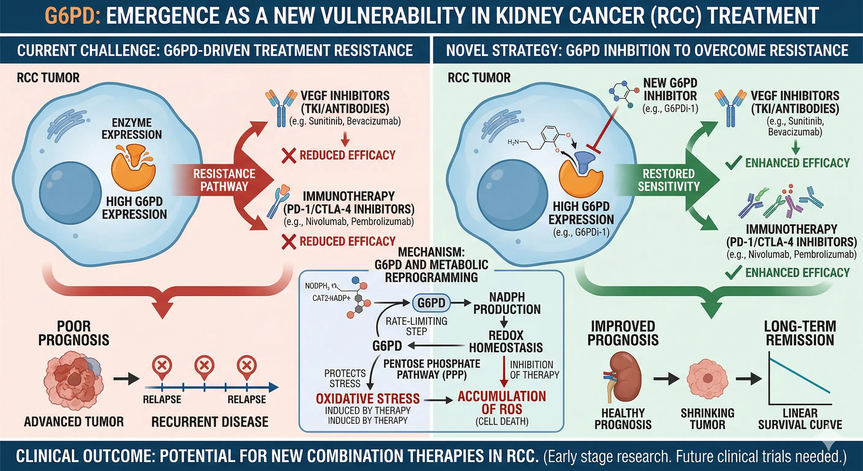
Inhibiting glucose-6-phosphate dehydrogenase (G6PD) produced different responses across renal cell carcinoma (RCC) cell lines. The researchers analyzed patient tissue and public gene-expression data, then compared drug responses in two RCC cell lines. The findings support the possibility that G6PD is a metabolic vulnerability in some tumors. They do not provide clinical evidence that targeting G6PD overcomes treatment resistance in patients or enhances immunotherapy.
RCC is treated with vascular endothelial growth factor receptor (VEGFR)-targeting drugs and immunotherapy, but treatment resistance limits long-term benefit. G6PD catalyzes the first step of the pentose phosphate pathway, which supplies precursors for nucleic acid synthesis and helps maintain cellular redox balance. The researchers examined whether G6PD expression was linked to tumor aggressiveness and whether inhibiting the enzyme reduced cancer-cell viability.
Key findings
The tissue analysis included 121 RCC cases and 12 adjacent non-tumor kidney tissue samples. G6PD immunostaining was negative in 80.2% of tumors, but papillary RCC had higher staining scores than clear-cell RCC. In clear-cell RCC, higher protein expression was associated with higher nuclear grade, tumors larger than 7 cm, and necrosis. The findings do not mean that G6PD is highly expressed in all RCCs.
The survival analysis of public gene-expression data included 530 clear-cell and 288 papillary RCC cases. In both subtypes, higher G6PD expression was associated with shorter overall survival. However, the researchers found no overall-survival difference by G6PD protein expression in their own tissue cohort. They suggested that this may have been due to the small number of metastatic cases and deaths.
The relationship with drug response was assessed using public cell-line data, not patient data. G6PD expression was 3.7 times as high in the lenvatinib nonresponder group as in the responder group (p=0.048). It was also 1.6 times as high in the cabozantinib nonresponder group, but the difference was not statistically significant (p=0.23). The expression difference was not supported to the same degree for both drugs, and this analysis did not establish the ability to predict patient treatment response.
In functional experiments, the researchers treated 786-O and ACHN cells with G6PDi-1, an experimental G6PD inhibitor. In 48-hour experiments at concentrations of 25–200 μM, the maximum reduction in the viability measure was 78% in 786-O cells and 36% in ACHN cells. However, this measure was based on metabolic activity, assessed by resazurin reduction, so the decrease cannot be interpreted directly as a cell-death rate. ACHN cells showed increased resazurin reduction at lower concentrations. The paper explains that this increase does not indicate increased proliferation and may reflect metabolic or redox adaptation.
Implications and outlook
Responses to G6PD inhibition differed across RCC models. In 786-O cells, suppression of viability and colony formation was pronounced, whereas ACHN cells were more sensitive to cabozantinib. In wound-closure assays assessing cell migration, cabozantinib had the strongest inhibitory effect. G6PDi-1 did not perform best in every assay.
The different test concentrations used for each drug should also be considered when comparing them. The tissue analysis was based on a single-region cohort, and the functional experiments were limited to two cell lines. The researchers did not directly analyze metabolic pathways, so the mechanisms behind the differing responses remain to be verified. G6PD is a candidate target for further study, but these findings alone are not sufficient to propose a patient-selection test or a strategy to replace existing treatments. The study was published in International Journal of Molecular Sciences. Full text of the paper
Renal cell carcinoma (RCC), the prevalent form of kidney cancer with rising global incidence, continues to challenge treatment efficacy despite advancements in VEGF-targeted tyrosine kinase inhibitors (TKIs) and immunotherapy. Persistent therapeutic resistance underscores the critical need for additional biomarkers indicative of tumor aggressiveness and novel therapeutic targets. This study investigates the role of glucose-6-phosphate dehydrogenase (G6PD), where altered expression has been implicated in various cancers, as a potential biomarker and therapeutic target in RCC management.
This study provides a starting point for follow-up research to distinguish which RCC models depend on G6PD. During drug development, researchers could compare G6PD expression, gene dependency, and inhibitor response to identify sensitive models. High expression alone does not establish that a patient's response to a drug can be predicted. Independent tissue cohorts are needed to re-examine the associations between expression and tumor size, grade, and prognosis. In vivo efficacy and safety, as well as appropriate dosing conditions, also need to be established; for now, G6PD is best viewed as a candidate research target rather than a treatment ready for patients.