Restoring Peptide Loading Complex Function: An Immunotherapeutic Strategy to Enhance Tumor Immune Visibility
Background
For immune checkpoint inhibitors and T-cell therapies to be effective, cancer cells must present tumor antigens on their cell surface. The major histocompatibility complex class I (MHC-I) plays a central role in this process. Cytotoxic CD8⁺ T cells recognize peptides presented by MHC-I to identify and eliminate abnormal cells. However, if cancer cells downregulate MHC-I expression or disrupt antigen processing, they become invisible to the immune system, rendering these therapies ineffective.
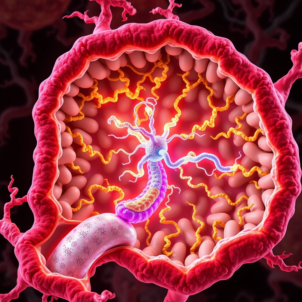
The peptide-loading complex (PLC), located in the endoplasmic reticulum (ER), is a molecular assembly that loads peptides generated in the cytoplasm onto MHC-I and performs quality control. It consists of the transporters associated with antigen processing (TAP1/TAP2), the adapter protein tapasin, and the chaperones calreticulin and ERp57, along with the MHC-I heavy chain and β2-microglobulin (β2M). In tumors, genetic alterations, as well as transcriptional, post-transcriptional, and epigenetic modifications, can impair the function of PLC components, leading to immune evasion and treatment resistance.
Key Findings
This review summarizes the evidence supporting the role of PLC dysfunction in cancer immune evasion and treatment resistance, focusing on the individual components of the complex. The authors argue that the PLC should be considered not just an MHC-I assembly helper, but a key regulator of tumor "immune visibility."
TAP1 and TAP2 transport peptides generated by the proteasome into the ER. Tapasin anchors MHC-I molecules near TAP, facilitating peptide loading and promoting the exchange of weakly bound peptides for more stable ones. Calreticulin and ERp57 assist in the folding and stabilization of MHC-I complexes, while β2M supports the transport of peptide-MHC-I complexes to the cell surface. Disruption of any of these components can lead to a reduction in stable MHC-I/peptide complexes, impairing recognition by cytotoxic T lymphocytes (CTLs).
The review also outlines four potential strategies for restoring PLC function: upregulation of antigen-presenting genes, including TAP and tapasin, using interferon-gamma (IFN-γ); gene therapy to supplement TAP1 or tapasin in cancer cells; epigenetic modulation using DNA methyltransferase inhibitors (DNMTi) and histone deacetylase inhibitors (HDACi); and targeting microRNAs that repress PLC transcription. These strategies differ from conventional immunotherapies, which primarily focus on blocking inhibitory signals on T cells, by aiming to reactivate antigen presentation machinery in cancer cells.
Significance and Outlook
The status of the PLC may serve as a biomarker to predict response to immune checkpoint inhibitors, cancer vaccines, tumor-infiltrating lymphocyte (TIL) therapy, and T-cell receptor-engineered T-cell therapy. Analyzing TAP1, TAP2, tapasin, β2M, and epigenetic modifications in tumor tissue, in addition to measuring MHC-I expression, could help identify the specific steps at which antigen presentation is failing. Reversible expression suppression may be amenable to treatment with IFN-γ or epigenetic modulators, while gene deletion or loss-of-function mutations may require gene supplementation or MHC-I-independent immunotherapies.
However, most of the proposed therapies are still in preclinical or early clinical stages. IFN-γ can induce both antigen presentation and immune-inhibitory factors such as PD-L1, and DNMTi and HDACi have broad targets, potentially leading to toxicity and unintended transcriptional changes. PLC defects may also vary among different cancer types, patients, and even within individual tumors, making it difficult to restore function with a single agent. Future research should focus on patient selection based on specific defect types, immune peptidome analysis to directly assess complex function, and clinical trials to evaluate the optimal combination and sequencing of PLC-targeted therapies with immune checkpoint inhibitors.
The peptide-loading complex (PLC) is a pivotal endoplasmic reticulum (ER) protein machinery that facilitates peptide translocation, editing, and loading onto major histocompatibility complex class I (MHC-I) to allow recognition of infected or transformed cells by CD8⁺ cytotoxic T lymphocytes (CTLs). PLC comprises the peptide transporter complex formed of transporters associated with antigen processing (TAP1/2), the adapter protein tapasin, chaperones such as calreticulin (CRT) and ERp57, and β2-microglobulin (β2M), ensuring the proper assembly of MHC-I/peptide complexes for subsequent antigen presentation on the cell surface. In cancer, tumorigenesis often involves structural malfunctions and transcriptional, post-transcriptional, and epigenetic modifications that impair the functioning of PLC proteins. This eventually causes loss of MHC-I cell-surface expression, failure of antigen presentation, and escape from CTL-mediated immunosurveillance. Although malfunctions in antigen-processing machineries are well known, herein we provide an overview of the PLC as a major determinant of tumor immune escape and a potential therapy target. In this regard, recent evidence indicates that individual PLC components not only affect MHC-I/antigen presentation but also influence tumor development and progression, immune evasion, and treatment resistance. Furthermore, we explore existing or evolving therapeutic approaches to recover/reprogram PLC functions such as IFN-γ-induced PLC activation, TAP1 and tapasin gene therapy, epigenetics-based treatments such as DNA methyltransferase inhibitors (DNMTi) and histone deacetylase inhibitors (HDACi), and microRNA targeting strategy.
In the clinic, PLC components in biopsy samples obtained before treatment with immune checkpoint inhibitors can be assessed at the genomic, transcriptomic, and proteomic levels to identify the causes of treatment resistance. For example, tumors with TAP1 and tapasin epigenetically silenced could be treated with DNMTi or HDACi followed by immune checkpoint inhibitors. Conversely, in cancers with complete loss of β2M, PLC activation may not be sufficient to restore MHC-I expression, and NK cell-based therapies or MHC-I-independent cell therapies may be more appropriate. Industrially, potential development candidates include PLC function assays combined with immune peptidome analysis for companion diagnostics, tumor-selective TAP1/tapasin delivery systems, and microRNA inhibitors.