Human Kidney Development Spatial Map Reconstructed from 700,000 Cells: Microenvironmental Signals Determine Cell Fate
Background
The kidney is an essential organ that filters blood waste and maintains fluid and electrolyte balance in the body. The nephron is the basic functional unit responsible for these functions. Humans are born with approximately 200,000 to 2 million nephrons. Nephrons are not newly formed after birth, and they cannot regenerate after damage. Consequently, the number of nephrons present at birth largely determines lifelong kidney health. Being born with a low number of nephrons significantly increases the risk of developing hypertension or chronic kidney disease (CKD) in adulthood.
Previous studies primarily focused on the genetic programs within kidney cells as the determinants of cell differentiation. However, analyzing individual cell genes alone makes it difficult to explain the actual location of cells within the tissue and how they interact with neighboring cells. Therefore, the specific mechanisms by which cells interact and form complex filtration structures during the early stages of kidney development remain largely unknown. To create therapeutic kidney tissue in the laboratory or prevent related diseases, it is necessary to first elucidate the spatiotemporal rules governing cell arrangement and fate determination during the embryonic stage.
Key Findings
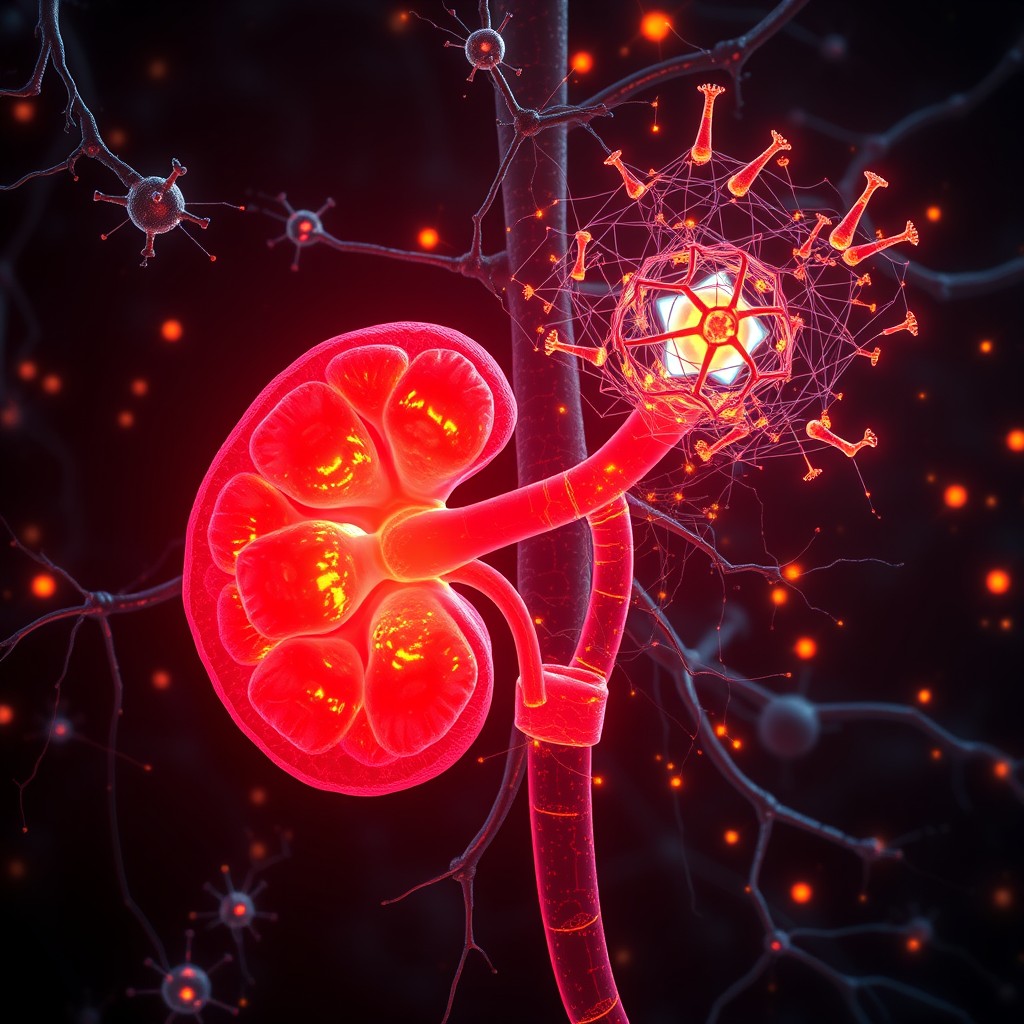
A joint research team from the University of Pennsylvania and the Children's Hospital of Philadelphia sought to solve this puzzle by combining single-cell RNA sequencing (scRNA-seq) and spatial transcriptomics techniques. The analysis included more than 700,000 human embryonic kidney cells. Based on this large dataset, the research team combined the gene expression status of individual cells with three-dimensional spatial information to create a detailed spatial map of kidney development.
The analysis revealed that the fate of kidney progenitor cells is more influenced by soluble signaling molecules secreted by neighboring cells than by internal genetic programs. Notably, insulin-like growth factor 2 (IGF2) was identified as a key signaling molecule that maintains the undifferentiated state of nephron progenitor cells (NPCs). If the IGF2 signal is blocked, the progenitor cells fail to differentiate into kidney cells and undergo apoptosis. This provides an important clue to explain why specific genetic variations associated with adult kidney size are located near the IGF2 gene.
The plasticity of cell differentiation was also newly discovered. Traditional developmental biology suggests that once a cell begins to differentiate into a specific lineage, its fate is fixed. In this study, progenitor cells that were already following the differentiation pathway of proximal tubule cells were observed to exhibit the ability to change their fate into other cell types within the renal corpuscle in response to changes in the surrounding microenvironmental signals. This demonstrates that cell fate determination is not a fixed process but rather a dynamic process in which cells interact with their surroundings and continuously explore new pathways.
Significance and Prospects
The kidney development spatial map elucidated in this study represents a milestone that will advance the technology of artificial organ fabrication. When creating kidney organoids in the laboratory using stem cells, mimicking the microenvironmental signals of actual kidney cells can enable the induction of highly functional artificial kidney tissue. By controlling the mechanisms that determine the number of nephrons at the molecular level, it is possible to develop new drugs to prevent nephron loss in premature infants or fetuses with impaired kidney development.
However, there are still challenges to be overcome before the results of this study can be applied in clinical practice. Recreating the complex microenvironmental signals of the embryonic developmental stage in an ex vivo environment is technically challenging. There is also a risk that multiple growth factors, including IGF2, may induce nonspecific cell proliferation, so further technical improvements are needed to precisely control the timing and concentration of signaling molecules. Future research is needed to conduct comparative analyses between animal models and actual clinical samples to verify the safety of the treatment.
Nature Genetics, Published online: 30 July 2026; doi:10.1038/s41588-026-02665-0The spatial organization of human kidney development is explored using single-cell RNA sequencing and spatial transcriptomics, highlighting the crucial role of microenvironmental signals in guiding cell fate.
This study is expected to be used directly as a tool for early screening of individuals at risk of chronic kidney disease and for developing treatment strategies in actual medical settings. Since the signaling pathways that regulate the number of nephrons during kidney formation have become clear, it is possible to identify indicators for predicting the risk of kidney developmental disorders during fetal gene screening in pregnant women. By analyzing genetic variations around IGF2, it is possible to identify high-risk individuals who are susceptible to chronic kidney disease in adulthood and to provide timely personalized lifestyle modifications or drug prescriptions.
In the field of artificial organs, it will be possible to produce more perfect kidney organoids. Existing kidney organoids have limitations in drug toxicity evaluation and development of organs for transplantation because they have poor vascular formation and immature nephron structure compared to actual organs. The 700,000-cell spatial map constructed in this study serves as a detailed recipe that tells us when and at what concentration to administer soluble substances when making organoids. As a result, it will be possible to mass-produce high-quality kidney models with filtration efficiency similar to that of actual kidneys, significantly reducing the time required for drug toxicity evaluation of new drug candidates.