Unlocking the Survival of Sleeping Sickness Parasites: Molecular Structure of the Mitochondrial RNA Editing Complex Revealed
Background
Trypanosoma brucei (T. brucei), the causative agent of African sleeping sickness, possesses a unique survival strategy. The mitochondrial genome of this parasite is defective, unable to encode complete proteins. For genetic information to be properly expressed, the transcribed messenger RNA (mRNA) must undergo a peculiar gene editing process involving the precise insertion or deletion of uridine (U) nucleotides. This process, guided by guide RNA (gRNA), forms the basis for synthesizing proteins necessary for the parasite's respiration and energy metabolism.
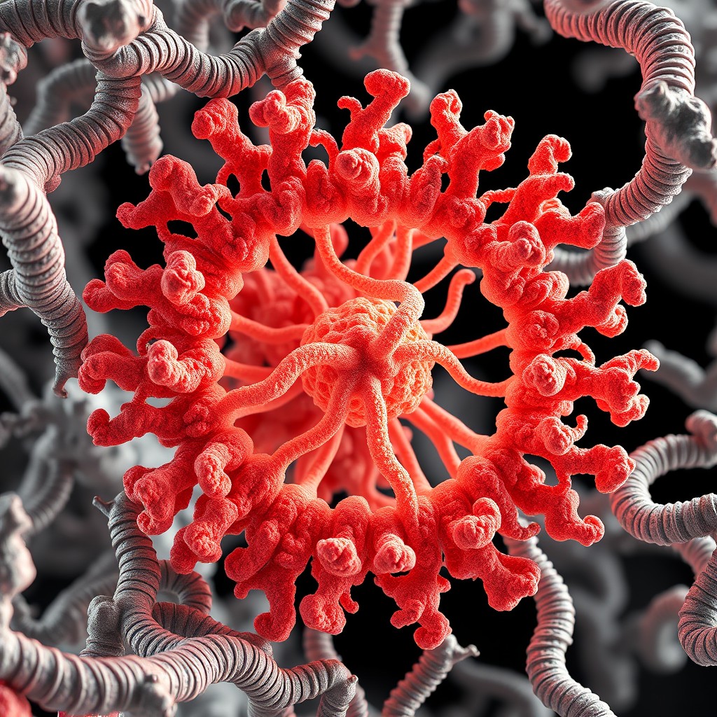
The large, multi-protein complex that drives this editing process is the RNA-Editing Catalytic Complex (RECC), also known as the editosome. The editosome has been a focus of attention in the molecular biology community for decades, but its large size, complexity, and heterogeneity have repeatedly hindered the determination of its detailed three-dimensional structure. The mechanism by which this complex recognizes the bound state of gRNA and mRNA and induces chemical reactions at precise locations has long remained a mystery. Previous low-resolution models could not clearly elucidate how the individual protein components interact, which has slowed the development of targeted therapeutics.
Key Findings
A research team led by Professor Ruslan Aphasizhev at the University of California, Los Angeles (UCLA) used cryo-electron microscopy (cryo-EM) to determine the three-dimensional structure of RECC1 and RECC2, the major complexes that make up the T. brucei editosome, at a resolution of up to 2.99 Ångströms. The high-resolution map obtained by the research team provides a detailed visualization of the architecture of the catalytic core, which has been shrouded in mystery.
The structural analysis revealed that the RECC1 complex, which mediates U deletion, consists of a KREN1 endonuclease dimer, KREPB8, and KREX1 exo-Uase, which removes U nucleotides, all intricately assembled and functioning in concert. These components tightly fix the double helix formed by gRNA and mRNA, and then sequentially perform reactions to expose, cleave, and delete specific U bases. In contrast, the RECC2 complex, which induces U insertion, is structurally organized around KREN2 endonuclease and KREPB7, forming an active site optimized for adding U bases to vacant spaces.
The research team compared the structural differences between the two complexes and demonstrated how opposing reactions, deletion and insertion, are precisely controlled on the same RNA strand. This study visualized the molecular mechanism of the editing cascade that occurs within the parasite's mitochondria, solving a long-standing molecular biology problem.
Significance and Prospects
This structural determination provides a blueprint for precisely targeting the molecular machinery unique to trypanosome parasites. Since human cells do not have a similar mitochondrial RNA editing system, substances that interfere with the catalytic core of the editosome could be safe drug candidates that selectively target parasites without harming humans. This principle aims to fundamentally block the mitochondrial activity essential for parasite survival.
However, there are also challenges to be overcome before this can lead to actual therapeutics. RNA editing is not a fixed, single-step process, but rather a non-processive reaction in which multiple RECC complexes repeatedly bind to and dissociate from the substrate RNA. The specific dynamic relationships and binding cycles that control these dynamic changes have not yet been fully elucidated. Therefore, subsequent research should involve real-time observation of the dynamic editing machinery in action and molecular dynamics analysis to identify the optimal timing for interfering with binding.
Nature, Published online: 22 July 2026; doi:10.1038/s41586-026-10831-xCryo-electron microscopy sheds light on the architecture of the Trypanosoma brucei mitochondrial editosome and reveals how this assembly mediates mRNA editing through the insertion and deletion of uridine nucleotides.
The high-resolution structural information of RECC1 and RECC2 revealed in this study will be directly utilized in structure-based drug design (SBDD) in the field of new drug development. The pharmaceutical industry can use the 3D coordinate information at 2.99 Å resolution to screen, using computer-aided virtual screening, for small molecule compounds that bind to the active site of KREN1 and KREN2 or the active pocket of KREX1, thereby inhibiting their enzymatic activity.
This approach can overcome the serious brain toxicity and inconvenience of injection associated with existing African sleeping sickness drugs, and can enable the development of a new generation of orally available, low-toxicity anti-trypanosomal drugs. Furthermore, this unique U insertion and deletion mechanism provides inspiration for designing artificial RNA editing tools for researchers in the field of synthetic biology, and also has the potential for technological expansion into the field of synthetic biology.